Proteins
A protein molecule is made up of a long chain of amino acids each linked to its neighbor through a covalent peptide bond. They are therefore referred to as polypeptides.
Amino Acids
Amino acids are the building blocks of proteins. The structure of amino acids consists of a central carbon atom that is bonded to:
one carboxylic acid group
one amino group
a side group (side-chain)
The side group is responsible for the uniqueness of an amino acid since it affects its:
shape
size
composition
pH
charge
A protein is universally accepted to have four levels of structural organization:
Primary Structure

The primary structure of proteins is the specific linear sequence of amino acids that make up the polypeptide chain.
It constitutes the covalent peptide bonds that join the amino acids together.
The sequence of the amino acids will ultimately decide the nature of the proteins function.
Secondary Structure
The secondary structure of proteins describes certain portions or stretches of the polypeptide chain and refers to the three-dimensional arrangement of atoms in the molecule.
What causes secondary structures?
Secondary structure is caused by the need for neutralization of the polar groups in the main chain via hydrogen bonds. This allows the hydrophobic regions of the protein to settle into the interior.
Constituents of secondary structures:
(1) Alpha Helices

Alpha helices (α-helices) are repeating, twisted spiral or coiled structures stabilized by hydrogen bonds that generally form between the carboxyl oxygen of each amino acid residue and amide nitrogen of the amino acid situated four residues away (i.e. they are perpendicular to the polypeptide chain). The side-chains radiate outward and are staggered to minimize steric hindrance.
The average length of an α-helix is approximately ten residues = three helical turns. α-helices are either left-handed or right-handedwith the latter being most often observed.
Net dipole moment is also for an α-helix is as follows:
partial positive at N-terminus
partial negative at C-terminus
This favors the bonding of negatively charged groups (such as phosphates, etc.)
α-helices are the major structural componentsin many proteins. Some globular proteins contain mostly α-helices connected by turns or loops. A few examples are:
Hemoglobin --> about 7% are α-helices
small DNA binding helices
membrane-spanning helices
amphipathic helices
coiled coils (super-secondary structures)
*An interesting feature of the side-chain of Proline that makes it bad for α-helices is that the side-chain is bonded to the N atom eliminating any chances of hydrogen bonding and makes the polypeptide chain rigid.
On the other hand, the small side-chain of Glycine generates a wide array of Ψ and Φ angles. Hence both Proline and Glycine are often found at the boundaries of α-helices and in turns.
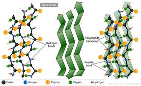
(2) Beta Sheets

Beta sheets (β-sheets) are segments of polypeptides that lie side by side. This causes the backbone of each segment to adopt a pleated conformation. Hydrogen bonds are formed between the carboxyl oxygen of one strand and the amide nitrogen of the adjacent strand (i.e. they are parallel to the sheets). Their highly extended form results in resistance to tensile forces.
The side-chains are perpendicular to the sheet and jut out on either side of the sheet giving each face a unique property (i.e. polarity)
β-sheets are typically found in the interior of proteins and are generally of three types:


parallel - adjacent β-strands run in same direction
anti-parallel - adjacent β-strands run in opposite direction
mixed
β-barrels are large β-sheets that are twisted and coiled in the form of a closed structure in which the hydrophobic side-chains line the outer surface to interact with the lipid bilayer.
**β-sheets cannot be transmembrane unless they are part of a β-barrel structure.
β-sandwiches are structures in which β-sheets are stacked upon one another; side chains are often hydrophobic and complementary.
(3) Hinges, Turns, Loops or Finger-like Extensions
These are the most flexible portions of the polypeptide chain and sites where the majority of biological activity takes place.
Super secondary structures
These are folding motifs composed of different combinations of secondary structures formed by interactions between the side-chains in order to solve the folding requirements by minimizing energy.
Motifs
Motifs are entities containing specific combinations of secondary structures having a particular topology. Some examples are:
coiled coils = alpha helices wrap around one another and form a structure that is thermodynamically stable; found in myosin
β-barrels = found in enzymes
zinc-finger motifs = DNA binding (found in transcription factors)
helix-turn-helix = DNA binding (found in transcription factors); calcium binding
Domains and Modules
Domains are parts of the polypeptide chain that are able to fold independently of one another into stable, compact substructures and usually key out a certain function.
Throughout the course of evolution, proteins with unique combinations of activities have been generated through shuffling of the domains.
Tertiary Structure
Tertiary structure of the protein describes the three-dimensional conformation of the entire polypeptide.
It is stabilized by non-covalent interactionsbetween the side-chains of proteins and leads a protein to attain the most thermodynamically stable conformation.
Quaternary Structure
Quaternary structure of a protein describes the relationship of the different polypeptide chainsthat make up a multiple subunit protein or a protein complex.


No comments:
Post a Comment